Read Pocket Medicine: The Massachusetts General Hospital Handbook of Internal Medicine Online
Authors: Marc Sabatine
Tags: #Medical, #Internal Medicine
Pocket Medicine: The Massachusetts General Hospital Handbook of Internal Medicine (132 page)
1
Parasternal long-axis view
allows visualization of the right ventricle (RV), ventricular septum (VS), posterior wall (PW) aortic valve cusps, left ventricle (LV), mitral valve, left atrium (LA), and ascending thoracic aorta (Ao). *Pulmonary artery. (Top: From
Mayo Clinic Proceedings
. [Tajik AJ, Seward JB, Hagler DJ,
et al.
Two-dimensional real-time ultrasonic imaging of the heart and great vessels: Technique, image orientation, structure identification, and validation.
Mayo Clinic Proceedings
, 1978;53:271–303], with permission. Bottom: From Oh JK, Seward JB, Tajik AJ.
The Echo Manual
,
3rd ed
. Philadelphia: Lippincott Williams & Wilkins, 2006. By permission of Mayo Foundation for Medical Education and Research. All rights reserved.)

2
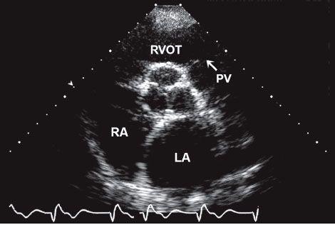
Parasternal short-axis view at the level of the aorta:
LA, left atrium; PV, pul-monary valve; RA, right atrium; RVOT, right ventricular outflow tract. (Top: From
Mayo Clinic Proceedings
. [Tajik AJ, Seward JB, Hagler DJ,
et al.
Two-dimensional real-time ultrasonic imaging of the heart and great vessels: Technique, image orientation, structure identification, and validation.
Mayo Clinic Proceedings
, 1978;53:271–303], with permission. Bottom: From Oh JK, Seward JB, Tajik AJ.
The Echo Manual
,
3rd ed
. Philadelphia: Lippincott Williams & Wilkins, 2006. By permission of Mayo Foundation for Medical Education and Research. All rights reserved.)
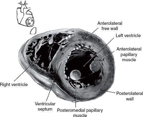

3 Parasternal short-axis view at the level of the papillary muscles:
AL, anterolateral papillary muscle; PM, posteromedial papillary muscle; RV, right ventricle; VS, ventricular septum; LV, left ventricle. (Top: From
Mayo Clinic Proceedings
. [Tajik AJ, Seward JB, Hagler DJ,
et al.
Two-dimensional real-time ultrasonic imaging of the heart and great vessels: Technique, image orientation, structure identification, and validation.
Mayo Clinic Proceedings
, 1978;53:271–303], with permission. Bottom: From Oh JK, Seward JB, Tajik AJ.
The Echo Manual
,
3rd ed
. Philadelphia: Lippincott Williams & Wilkins, 2006. By permission of Mayo Foundation for Medical Education and Research. All rights reserved.)


4
Apical four-chamber view:
Note that at some institutions the image is re-versed so that the left side of the heart appears on the right side of the screen. LA, left atrium; LV, left ventricle; RA, right atrium; RV, right ventricle. (Top: From
Mayo Clinic Proceedings
. [Tajik AJ, Seward JB, Hagler DJ,
et al.
Two-dimensional real-time ultrasonic imaging of the heart and great vessels: Technique, image orientation, structure identification, and validation.
Mayo Clinic Proceedings
, 1978;53:271–303], with permission. Bottom: From Oh JK, Seward JB, Tajik AJ.
The Echo Manual
,
3rd ed
. Philadelphia: Lippincott Williams & Wilkins, 2006. By permission of Mayo Foundation for Medical Education and Research. All rights reserved.)
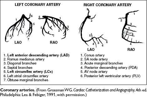
Coronary Angiography
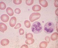
Peripheral Blood Smears

1
Normal smear.

2
Hypochromic, microcytic anemia due to iron-deficiency.
3
Macrocytic anemia due to pernicious anemia; note macro-ovalocytes and hypersegmented neutrophils.

Other books
Watching Sin (A Fetish and Fantasy Short Story) by Lori King
Harriet Doerr by The Tiger in the Grass
Owned By The Alphas: Part Two by Faleena Hopkins
The Red Pavilion by Jean Chapman
Autumn Falls by Bella Thorne
Sinful Secrets: Take Me (Billionaire Breeding Erotic Romance) by Wade, Natasha
Serious Sweet by A.L. Kennedy
Pieces of You by J F Elferdink
Darned if You Do by Monica Ferris
The Passionate and the Proud by Vanessa Royall