Read Breast Imaging: A Core Review Online
Authors: Biren A. Shah,Sabala Mandava
Tags: #Medical, #Radiology; Radiotherapy & Nuclear Medicine, #Radiology & Nuclear Medicine
Breast Imaging: A Core Review (12 page)
A. Approximately 10% of all breast cancers occur in the subareolar location.
B. Breast cancers in the subareolar location are more common in women than in men.
C. Breast cancers in the subareolar region are easy to detect, due to a lack of superimposed breast tissue in this location.
D. Breast cancers in this location are associated with earlier lymphatic spread via the retroareolar Sappey plexus.
19
A 55-year-old female presents for a screening mammogram. A mass is detected, which after further evaluation and biopsy proves to be invasive ductal carcinoma. What percentage of breast cancers in females is detected in this location?
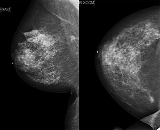
A. 7%
B. 17%
C. 27%
D. 37%
20
A 60-year-old female presents for a mammogram. A mass is detected at the site of the palpable abnormality in the upper outer quadrant. What percentage of breast cancers in females occurs in the upper outer quadrant?
A. 21%
B. 41%
C. 61%
D. 81%
21
A 29-year-old female presents with a palpable abnormality in the right breast. Given the ultrasound image below, what is the most likely diagnosis?
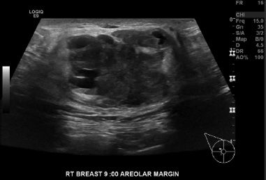
A. Galactocele
B. Lipoma
C. Hamartoma
D. Lymph node
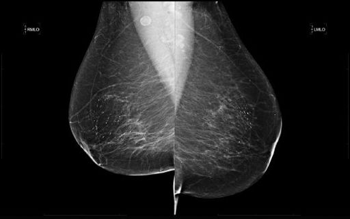
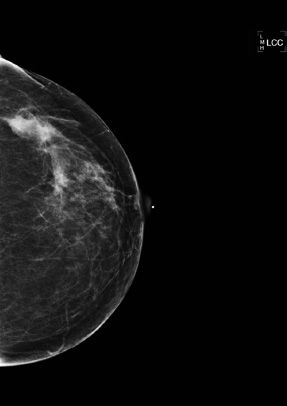
22
A 56-year-old female presents for a baseline screening mammogram. What is the appropriate BI-RADS classification?
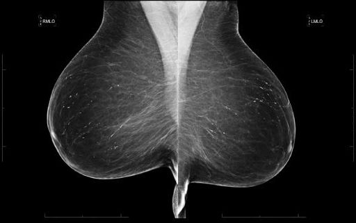
A. BI-RADS 0
B. BI-RADS 2
C. BI-RADS 3
D. BI-RADS 4A
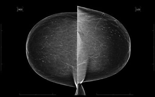
23
A 62-year-old female presents for an annual screening mammogram.
23a
Based on the images, what is the BI-RADS Category assessment?
A. BI-RADS 0
B. BI-RADS 2
C. BI-RADS 3
D. BI-RADS 4
23b
Based on the images, what is the recommendation?
A. Incomplete—Needs additional workup
B. Benign—Recommend annual screening in 1 year
C. Probably benign—Follow-up in 6 months
D. Suspicious—Recommend biopsy under stereotactic guidance.
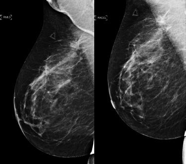
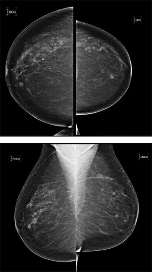
24a
Mammograms from 2011 are on the left and those from 2012 are on the right. The first pair represents CC projections, and the second pair represents MLO projections. The images are magnified to show the area of interest. What is the name of the radiologic sign that these images demonstrate?
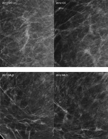
A. Cord sign
B. Cluster sign
C. Mirror sign
D. Tattoo sign
24b
What type of calcifications is represented in the above images?
A. Secretory calcifications
B. Fibroadenoma calcifications
C. Dermal calcifications
D. Milk of calcium calcifications
E. Fat necrosis calcifications
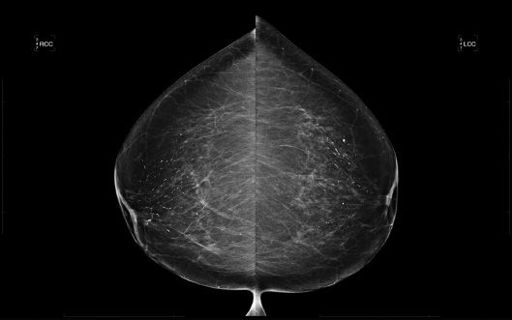
25
A 45-year-old asymptomatic female presents for a screening mammogram. craniocaudal (CC) and mediolateral oblique (MLO) views are shown below:
What BI-RADS assessment would you give?
A. BI-RADS 0
B. BI-RADS 2
C. BI-RADS 3
D. BI-RADS 4
26
Based on the location of the lesion in the left breast shown below, how do you expect the lesion to shift on a mediolateral (ML) view?
Other books
To Kill a Grey Man by D C Stansfield
The End of Eve by Ariel Gore
Blood Passage by McCann, Michael J.
Forbidden Love by Manro, Kaye
B006K5TA1E EBOK by Collins, Yvonne, Rideout, Sandy
The Zeppelin Jihad by S.G. Schvercraft
Lost and Found Family by Leigh Riker
Attica by Kilworth, Garry
Rebel Mechanics by Shanna Swendson
Serial Killer Investigations by Colin Wilson