Breast Imaging: A Core Review (16 page)
Read Breast Imaging: A Core Review Online
Authors: Biren A. Shah,Sabala Mandava
Tags: #Medical, #Radiology; Radiotherapy & Nuclear Medicine, #Radiology & Nuclear Medicine
References: Gunhan-Bilgen I, et al. Inflammatory breast carcinoma: Mammographic, ultrasonographic, clinical and pathologic findings in 142 cases.
Radiology
2002;223:829–838.
Kushwaha AC, et al. Primary inflammatory carcinoma of the breast. Retrospective review of radiological findings.
AJR Am J Roentgenol
2000;174:535–538.
30
Answer C.
There is whole-body radiation exposure from BSGI, with greatest effect on the bowel wall. BSGI has a lifetime attributable risk of mortality that is ~20 to 30 times greater than that of a complete screening digital mammogram. The density of breast tissue does not affect sensitivity, and BSGI is equally sensitive in dense and fatty breasts.
References: Brem RF, Rechtman LR. Nuclear medicine imaging of the breast: A novel, physiologic approach to breast cancer detection and diagnosis.
Radiol Clin North Am
2010;48:1055–1074.
Hendrick RE. Radiation does and cancer risks from breast imaging studies.
Radiology
2010;257:246–253.
31

Reference: Ikeda D.
Breast Imaging: The Requisites
. 2nd ed. St. Louis, MO: Elsevier Mosby; 2011:151.
32
Answer B.
If a lesion is visible only on mediolateral oblique (MLO) and true lateral views, the triangulation method is used to locate the lesion on the craniocaudal (CC) view. With the MLO view in the middle, a line drawn through the lesion in the MLO and true lateral views and extending through to the CC view will intersect lesion location on the CC view.
Reference: Berg WA, Birdwell R, Gombos EC, et al.
Diagnostic Imaging: Breast
. Salt Lake City, UT: Amirsys; 2006;II:0–13.
33
Answer B.
Mammography is the first imaging test of choice for a clinically suspicious mass in a male. A palpable mass that is occult or incompletely imaged on mammography warrants a targeted ultrasound.
Reference: Nguyen C, Kettler MD, Swirsky ME, et al. Male breast disease: Pictorial review with radiologic-pathologic correlation.
Radiographics
2013;33(3):763.
34
Answer C.
First-degree relatives include mother, father, sister, and daughter. Second-degree relatives include grandmother, aunt, and niece.
Reference: Berg WA, Birdwell R, Gombos EC, et al.
Diagnostic Imaging: Breast
. Salt Lake City, UT: Amirsys; 2006;II:0–24.
35
Answer D.
Peak incidence of breast cancer in these patients is at 15 years after treatment. They have an increased risk if radiation exposure is before 30 years of age. Preferred treatment is mastectomy with chemotherapy. Radiation is contraindicated.
References: Alm El-Din MA, Hughes KS, Raad RA, et al. Clinical outcome of breast cancer occurring after treatment for Hodgkin’s lymphoma: case control analysis.
Radiat Oncol
2009;4:19.
Berg WA, Birdwell R, Gombos EC, et al.
Diagnostic Imaging: Breast
. Salt Lake City, UT: Amirsys; 2006;IV:4-58.
36
Answer C.
Risk factors for breast cancer include early menarche, late menopause, nulliparous, atypical ductal hyperplasia (ADH), lobular carcinoma in situ (LCIS), personal history of breast cancer, first-degree relative with breast cancer, first birth after age 30, BRCA1 and BRCA2, radiation exposure at a young age.
Reference: Ikeda D.
Breast Imaging: The Requisites.
2nd ed. St. Louis, MO: Elsevier Mosby; 2011:24–25.
37
Answer D.
Interval cancers are defined as breast cancers presenting with chemical findings during the interval between recommended screenings. They can be mammographically occult or missed on prior mammography. Usually presenting as a new palpable lump compared to screen-detected cancers, there is an increased incidence of lobular and mucinous histology. There is a lower rate of ductal carcinoma in situ (DCIS). Women with very dense breasts have a higher incidence than those with fatty breasts. Prognosis for interval cancers is similar to symptomatic, unscreened breast cancers.
References: Berg WA, Birdwell R, Gombos EC, et al.
Diagnostic Imaging: Breast
. Salt Lake City, UT: Amirysis Inc; 2006;IV:2:140–143.
Buist DS, et al. Factors contributing to mammography failure in women aged 40–49 years.
J Natl Cancer Inst
2004;96:1432–1440.
Ikeda DM, et al. Analysis of 172 subtle findings on prior normal mammograms in women with breast cancer detected at follow up screening.
Radiology
2003;226:494–503.
38
Answer B.
The nipple should be seen on profile in at least one view to assess the subareolar area.
Reference: Bassett L, Hirbawi I, DeBruhl N, et al. Mammographic positioning: Evaluation from the viewbox.
Radiology
1993;188:803–806.
39
Answer C.
Adequate compression when obtaining mammograms is important for a number of reasons. It prevents motion, reduces scatter and spreads out the tissues better. It reduces the amount of radiation needed. Compression is usually less painful during the first half of the menstrual cycle and if the compression is applied gradually.
Reference: Berg WA, Birdwell R, Gombos EC, et al.
Diagnostic Imaging: Breast
. Salt Lake City, UT: Amirsis Inc.; 2006:I1:0-2–I1:0-3.
| 3 | Diagnostic Breast Imaging, Breast Pathology, and Breast Imaging Findings |
QUESTIONS
1
Based on this image, what is the most likely diagnosis?

A. Radial fold
B. Capsular contracture
C. Intracapsular rupture
D. Extracapsular rupture
2
What is the most common location for an intramammary lymph node?
A. Upper outer quadrant
B. Upper inner quadrant
C. Lower outer quadrant
D. Lower inner quadrant
3a
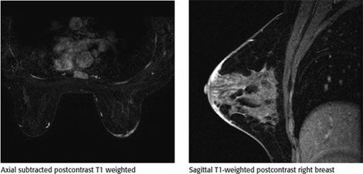
Based on the following images, the dominant finding is

A. Subareolar region nonmass-like enhancement
B. Enhancement of the pectoralis muscle
C. Unilateral skin thickening
D. Architectural distortion in the superior right breast
3b
What would be an appropriate differential diagnosis for the previous finding?
A. Related to phase of menstrual cycle
B. Mastitis
C. Hormone therapy
D. Renal failure
4a
A 16-year-old female presents with a palpable finding in her right breast. What is the most appropriate imaging test?
A. Unilateral right mammogram
B. Bilateral mammogram
C. Unilateral right ultrasound
D. Bilateral ultrasound
E. Unilateral right mammogram and ultrasound
4b
Which of the following statements regarding fibroadenomas is correct?
A. Giant fibroadenomas are more common in the Asian population.
B. Most fibroadenomas in teenagers are adult type.
C. Fibroadenomas are more common in postmenopausal women.
D. Fibroadenomas can be found equally in males and females.
4c
Based on the following image, what would be the most likely diagnosis?

A. Fat necrosis
B. Lymph node
C. Hematoma
D. Juvenile fibroadenoma
5
A 49-year-old female with no history of prior breast concerns or a family history of breast cancer presents with new onset right bloody nipple discharge. Based on the ultrasound images below, what is the most likely diagnosis?
Other books
Thunderland by Brandon Massey
Viaje al centro de la Tierra by Julio Verne
No Trace by Barry Maitland
The Remembered by Lorenzo, EH
Captain Future 13 - The Face of the Deep (Winter 1943) by Edmond Hamilton
Harry Truman by Margaret Truman
The Grafton Girls by Annie Groves
Acts of God by Mary Morris