Breast Imaging: A Core Review (33 page)
Read Breast Imaging: A Core Review Online
Authors: Biren A. Shah,Sabala Mandava
Tags: #Medical, #Radiology; Radiotherapy & Nuclear Medicine, #Radiology & Nuclear Medicine

References: Macura KJ, Ouwerkerk R, Jacobs MA, et al. Patterns of enhancement on breast MR images: interpretation and imaging pitfalls.
Radiographics
2006;26:1719–1734.
Liberman L, Morris, eds.
Breast MRI Diagnosis and Intervention
. New York, NY: Springer Science; 2005:238–265.
79
Answer C.
Invasive ductal carcinoma of the breast not otherwise specified is the most common type of invasive breast cancer to occur in pregnant and postpartum patients. A few other cancers have been reported but are significantly less frequent.
References: Sabate JM, Clotet M. Torrubia S, et al. Radiographic evaluation of breast disorders related to pregnancy and lactation.
Radiographics
2007;27:S101–S124.
Yang WT, Dryden MJ, Gwyn K, et al. Imaging of breast cancer diagnosed and treated with chemotherapy during pregnancy.
Radiology
2006;239:52–60.
Ahn BY, Kim HH, Moon WK, et al. Pregnancy and lactation-associated breast cancer: Mammographic and sonographic findings.
J Ultrasound Med
2003;22:491–497.
80
Answer D.
The breast MRI images demonstrate a mass in the left upper outer quadrant at posterior depth and a mass located in the left lower inner quadrant at middle depth, which is consistent for multicentric disease.
Multicentric disease
refers to more than one lesion in more than one quadrant of the breast.
Multifocal disease
refers to more than one lesion in a single quadrant of the breast. Since the masses are 3 to 4 cm on opposite sides of the breast, this patient would likely be treated with a mastectomy. Contraindications to whole-breast radiation therapy are
• Pregnancy
• Previous breast radiation
• Multicentric or diffuse disease
• Collagen vascular disease
• Poor cosmetic result (relative contraindication)
Reference: Ikeda D.
Breast Imaging: The Requisites
. 2nd ed. St. Louis, MO: Elsevier Mosby; 2011:301.
81a
Answer B.
81b
Answer B.
A galactocele is typically seen in lactating or postlactating women. A galactocele is a focal collection of breast milk. On mammography, a galactocele appears as a low- or equal-density mass with a pathognomonic fat-fluid appearance on the horizontal beam imaging (lateral/medial or medial/lateral view). On ultrasound, there can be varying appearances depending on the content of fluid and solid milk components. Pathognomonic appearance on ultrasound is a fluid debris level with the fat rising on top of the galactocele and the milk/fluid layering dependently below. Galactoceles usually resolve spontaneously within a few weeks to months. Aspiration is usually therapeutic.
References: Ikeda D.
Breast Imaging: The Requisites
. 2nd ed. St. Louis, MO: Elsevier Mosby; 2011:379–380.
Shah BA, Fundaro GM, Mandava S.
Breast Imaging Review: A Quick Guide to Essential Diagnoses
. 1st ed. New York, NY: Springer; 2010:25–27.
82
Answer A.
A simple breast cyst is fluid collection with an epithelial lining. It is the most common breast mass in women. Simple cysts usually have a round, oval, or lobular appearance on mammography but cannot be distinguished from a solid mass without use of ultrasound. On ultrasound, a simple cyst is by definition a mass that is anechoic with imperceptible walls, a sharp back wall, through transmission, and enhanced posterior transmission of sound. A simple cyst can be left alone or can be aspirated if symptomatic.
References: Ikeda D.
Breast Imaging: The Requisites
. 2nd ed. St. Louis, MO: Elsevier Mosby; 2011:137–139.
Shah BA, Fundaro GM, Mandava
S. Breast Imaging Review: A Quick Guide to Essential Diagnoses
. 1st ed. New York, NY: Springer; 2010:80–81.
83
Answer D.
Several published studies have reported on the mammographic appearance of pregnancy-associated breast cancer, and the most common mammographic finding was a mass with or without calcifications followed by calcifications alone or associated with other findings such as architectural distortion or diffuse increase in breast density.
References: Sabate JM, Clotet M. Torrubia S, et al. Radiographic evaluation of breast disorders related to pregnancy and lactation.
Radiographics
. 2007;27:S101–S124.
84
Answer C.
Calcifications in axillary lymph nodes can be due to many reasons. Differential diagnosis includes granulomatous disease and metastatic disease from breast cancer or ovarian cancer. New development of calcifications in axillary lymph nodes should be biopsied to exclude metastatic disease from breast or ovarian cancer.
References: Dunnington GL, Pearce J, Sherrod A, et al. Breast carcinoma presenting as mammographic microcalcifications in axillary lymph nodes.
Breast Dis
1995;8:193–198.
Ikeda D.
Breast Imaging: The Requisites
. 2nd ed. St. Louis, MO: Elsevier Mosby; 2011:396.
85
Answer C.
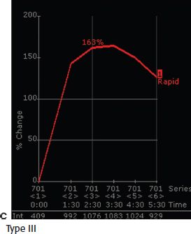
The breast MRI image shows an irregular breast mass with spiculated margins. If a mass has clear malignant features on morphology assessment, the mass should be biopsied irrespective of a benign kinetic curve.
Reference: Morris EA, Liberman L.
Breast MRI: Diagnosis and Intervention
. New York, NY: Springer; 2005:99.
86
Answer B.
Ultrasound images show a superficial oval complicated cystic mass with posterior acoustic enhancement along with a hypoechoic structure extending from the mass to the skin that is characteristic for either a sebaceous cyst (arising from a sebaceous gland) or an epidermal inclusion cyst (obstructed hair follicle). Clinically and on imaging, sebaceous cysts and epidermal inclusion cysts are indistinguishable from each other and usually require no treatment. Excision may be warranted if persistently painful or enlarge. There is no malignant potential for sebaceous cysts, and it is extremely rare in epidermal inclusion cysts. Therefore, a BI-RADS 2 category is the appropriate assessment following diagnostic workup.
Reference: Shah BA, Fundaro GM, Mandava S.
Breast Imaging Review: A Quick Guide to Essential Diagnoses
. 1st ed. New York, NY: Springer; 2010:188–189.
87
Answer B.
There has been interval decrease in size of both breasts on the current screening mammogram as compared to the 2-year prior screening mammogram. There are swirled areas of architectural distortion in the lower breast bilaterally on the MLO views and centrally on the CC views. There is an oval radiolucent mass in the lower inner right breast that represents fat necrosis. Findings are consistent for bilateral reduction mammoplasty.
Other characteristic mammogram findings seen are
• Elevation of the nipple with more skin inferior than superior
• Redistribution of fibroglandular tissue from upper outer quadrant to lower inner quadrant
Fat necrosis can be an associated finding seen in reduction mammoplasty.
Reference: Shah BA, Fundaro GM, Mandava S.
Breast Imaging Review: A Quick Guide to Essential Diagnoses
. 1st ed. New York, NY: Springer; 2010:49–50.
88
Answer C.
The images shown are that of bilateral subpectoral saline breast implants. There is a normal subpectoral saline implant in the left breast. When a saline breast implant ruptures, the saline disperses in the breast tissues and the envelope/shell of the implant shrinks back against the chest wall. Radial fold is infolding of the implant envelope/shell that is a normal finding seen in silicone breast implants. Radial folds appear as dark lines on MRI that extend to the periphery of the implant. Capsular contracture occurs when normal scar tissue forms a capsule around the breast implant and tightens/squeezes the breast implant. Capsular contracture may occur several months to years and can result in change in the breast shape, sensation of hardness of the breast, and/or breast pain.
References: Ikeda D.
Breast Imaging: The Requisites
. 2nd ed. St. Louis, MO: Elsevier Mosby; 2011:341.
Shah BA, Fundaro GM, Mandava S.
Breast Imaging Review: A Quick Guide to Essential Diagnoses
. 1st ed. New York, NY: Springer; 2010:236–237.
89
Answer C.
Mondor’s disease is a focal thrombophlebitis of a superficial vein in a breast. It can clinically present as a palpable cord–like mass with associated pain, tenderness, and erythema. Mondor’s disease is a rare condition that can be associated with trauma, breast surgery, and extreme physical activity. Treatment is not needed as it is self-limited, and the palpable finding resolves in 2 to 12 weeks. Supportive care is the appropriate treatment.
On mammography, Mondor’s disease can have negative mammogram or rarely a long tubular structure corresponding to the thrombosed vein.
On ultrasound, Mondor’s disease appears as a noncompressible tubular superficial structure with or without color Doppler flow depending on the degree of recanalization.
Reference: Ikeda D.
Breast Imaging: The Requisites
. 2nd ed. St. Louis, MO: Elsevier Mosby; 2011:399–400.
90
Answer A.
Classic features of neurofibromatosis type 1 (NF 1), also known as von Recklinghausen disease, have neurofibromas and café au lait spots. NF 1 is associated with neoplasms such as meningiomas, optic gliomas, neurofibrosarcomas, and pheochromocytomas. Neurofibromas can project over the breast and have an appearance of masses within the breast on mammography. However, a skin lesion can be differentiated from a mass in the breast by looking for a radiolucent halo around the mass.
Reference: Shah BA, Fundaro GM, Mandava S.
Breast Imaging Review: A Quick Guide to Essential Diagnoses
. 1st ed. New York, NY: Springer; 2010:41–42.
91
Answer D.
Poland syndrome is either congenital unilateral hypoplasia or absence of the pectoralis major muscle.
Poland syndrome is associated with increased incidence of:
• Breast cancer
• Leukemia
• Non-Hodgkin lymphoma
• Lung cancer
Reference: Shah BA, Fundaro GM, Mandava S.
Breast Imaging Review: A Quick Guide to Essential Diagnoses
. 1st ed. New York, NY: Springer; 2010:82–83.
Other books
Mental Floss: Instant Knowledge by Editors of Mental Floss
The Haters by Jesse Andrews
Supergirl by Norma Fox Mazer
The Alternative Hero by Tim Thornton
At the Highlander's Mercy by Terri Brisbin
The Jersey Vignettes by Bethany-Kris
Cold Sacrifice by Leigh Russell
Who Asked You? by Terry McMillan
The Wrong Kind of Money by Birmingham, Stephen;