Breast Imaging: A Core Review (42 page)
Read Breast Imaging: A Core Review Online
Authors: Biren A. Shah,Sabala Mandava
Tags: #Medical, #Radiology; Radiotherapy & Nuclear Medicine, #Radiology & Nuclear Medicine
E. 4.0%
35
What instrument is recommended by the ACR to determine focal spot size?
A. Phototimer
B. Densitometer
C. SMPTE pattern
D. Slit camera
E. Star-shaped camera
36
What is the mammography phantom used for in digital imaging?
A. Daily testing of the AEC
B. Weekly testing the anode
C. Daily checking of the focal spot
D. Weekly testing of system resolution
E. Monthly checking image contrast
37
The mammography tube has a window that consists of:
A. Beryllium
B. Molybdenum
C. Tungsten
D. Rhodium
E. Pyrex glass
38
In mammography, the cathode side of the tube should be placed by the:
A. Nipple
B. Chest wall
C. Lateral aspect of breast
D. Medial aspect of breast
E. Adipose tissue
39
What is the size of the large focal spot used for standard mammography?
A. 0.1 mm
B. 0.3 mm
C. 0.7 mm
D. 1.0 mm
40
Why does mammography utilize a lower peak kilovoltage than conventional radiography?
A. To improve soft-tissue contrast
B. To reduce the mean glandular dose received by the patient
C. To decrease exposure time
D. To improve scatter rejection
41
Which of the following is correct regarding the use of compression in mammography?
A. Increases image contrast and increased scatter radiation
B. Reduces breast thickness and requires a higher radiation dose
C. Reduces scatter radiation and decreases image contrast
D. Requires less kVp values and decreases motion blur
E. Spreads breast tissue to reduce superimposition and increase structural mottle
42
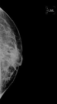
Below is a mammographic view taken at 28 kVp, 42 mAs, 3.1-cm thickness. Altering the parameters to 26 kVp, 110 mAs, and 3.1-cm thickness leads to which one of the following changes?

A. Shorter exposure time
B. Increased spatial resolution
C. Decreased glandular dose
D. Increased contrast
E. Decrease density
ANSWERS AND EXPLANATIONS
1
Answer A.
Grids are used routinely in mammography to increase image contrast. Most mammography systems have a moving grid with a ratio of 4:1 to 5:1 focused to the source to image distance. Grids do not compromise spatial resolution, but they do increase patient dose. However, the dose is still acceptably low, and the improvement in contrast is significant.
Reference: Bushong SC.
Radiologic Science for Technologists: Physics and Protection
. 10th ed. St. Louis, MO: Elsevier Mosby; 2013:381.
2
Answer C.
Breast compression results in decreased tissue thickness. The scatter to primary ratio for a compressed breast is 0.4 to 0.5, whereas the scatter to primary ratio for a noncompressed breast is 0.8 to 1.0. Reducing tissue thickness allows for use of lower mAs, which results in decreased radiation dose. Compression results in reduced exposure dynamic range because tissue is spread out creating a more uniform thickness. Magnification can be produced with an air gap.
Reference: Bushberg JT, Seibert JA, Leidholt EM, et al.
The Essential Physics of Medical Imaging
. 2nd ed. Philadelphia, PA: Lippincott Williams & Wilkins; 2001:207.
3
Answer C.
A very good, short white paper on mammography reading room design is Albert Xthona, “Designing the Perfect Reading Room for Digital Mammography,” Barco White Paper, 2003 and is available online at this link:
http://www.barco.com/barcoview/downloads/The_perfect_mammography_reading_room_2011_-_White_paper.pdf
. This is the primary resource for this question. The paper shows a long narrow room with all workstations along one wall, emphasizing the need to reduce light falling from one monitor to another. Thus, all the workstations are in a straight line.
A.
Though workstations are sometimes placed angled slightly toward each other; this greatly increases the background lighting at each monitor and substantially decreases the image contrast.
B.
Walls should be dark colored to reduce the ambient light. Light for walking safely should be very near the ground, with low-level lamps placed below the desks and pointing toward the floor.
D.
Mammography view boxes are sometimes used as a source of light for writing or moving about the room. This is not a good idea, and the reference shows how to better accommodate these needs.
4
Answer D.
This covers the range of target/filter combinations used in digital mammography, and the tungsten target + silver filter (k edge energy of 25 keV) will have the highest energy and greatest penetrating power.
Reference: Huda W.
Review of Radiographic Physics
. 3rd ed. Baltimore, MD: Lippincott Williams & Wilkins; 2010:51–52.
5
Answer E.
Moving grids (Bushong, p. 200ff) are used in contact mammography (but not in magnification mammography) solely for the reduction of scatter to the image receptor. Note that the Mammography Quality Safety Act (MQSA)
requires
[900.12(b)(4)] that
1 (ii) Systems using screen-film image receptors shall be equipped with moving grids matched to all image receptor sizes provided.
2 (iii) Systems used for magnification procedures shall be capable of operation with the grid removed from between the source and image receptor.
Grids are generally used for the same reasons for machines with digital receptors as well. Breast compression has many benefits
including
the reduction of scatter (Bushong, p. 380).
Answer choice A is wrong because “compression results in thinner tissue and therefore less scatter radiation (Bushong, p. 380). ” Scatter is higher at higher kV (Bushong, pp. 187, 188) and so answer choice B is wrong. Answer choice C is a little tricky. Breast compression reduces the scatter and thus the benefit of scatter reduction using a grid, but, in that, “the use of grids during (contact) mammography is routine (Bushong, p. 381),” the grid still provides enough benefit to warrant its use and so answer choice C is wrong. This subject is explored further in Gennaro et al. for magnification mammography; however, the air gap sufficiently reduces the effects of scatter so that the grid is not used (Bushberg et al., p. 210) and so answer choice D is incorrect.
References: Bushong SC.
Radiologic Science for Technologists
. 10th ed. St. Louis, MO: Elsevier; 2013.
Bushberg JT, Seibert JA, Leidholdt EM, et al.
The Essential Physics of Medical Imaging
. 2nd ed. Philadelphia, PA: Lippincott Williams & Wilkins; 2001.
Gennaro G, Katz L, Souchay H, et al. Grid removal and impact on population dose in full-field digital mammography.
Med Phys
2007;34(2):547–555.
MQSA:
http://www.fda.gov/Radiation-EmittingProducts/MammographyQualityStandardsActand Program/Regulations/ucm110906.htm
6
Answer C.
Many of the points in the discussion below are taken from an excellent tutorial on mammographic displays by Ehsan Samei.
Many of the quantitative terms (and there
are
many) used in optics have evolved over centuries of use and have a quaint, even charming, but often confusing feel to them.
Luminance
and
illuminance
are two of these terms. While both now have official SI definitions, they still bear a relationship to their everyday English meanings.
Luminance
is the perceived brightness of a display and can apply to both view boxes and the monitors used for digital display. (
Illuminance
describes the outside light falling on the display and while illuminance is good when reading a book, it degrades the images from monitors used for mammographic displays by reducing the perceived contrast.) Now on to the question.
The answer choice A is wrong because there is a limit to the brightness range comfortable (and at extremes, safe) for the human vision system. (That is why we have sunglasses, for example.) Answer choice B is somewhat better but again, as we go too high in the contrast ratio displayed, we again reach the limits of the adaption capabilities of the human visual system. We also run into problems due to the contrast reduction processes of veiling glare and reflection. The answer choice D is incorrect because even the best mammographic film cannot compete with the dynamic range in contrast of modern digital displays. (Film still beats digital displays in resolution, however. That is it can see tiny objects of high contrast better than digital displays.)
Answer choice C is correct. Modern digital displays can easily, as previously stated, exceed the contrast display capabilities of film so a good minimum contrast ratio is that of film on a standard view box. However, it should not be too high for the reasons given above in discussing answer choice B.
Reference: Samei E. Technological and psychophysical considerations for digital mammographic displays.
Radiographics
2005;25:491–501.
7
Answer A.
Imagine looking at a display with a uniform background of brightness
L
. The display is divided into two sections but initially both are matched in brightness so that they appear as one. Now imagine that one side (e.g., the left side) is
very
slowly made brighter while you are viewing the display. At some point you perceive that there are two sections with the left half just barely brighter than the right half. This difference in brightness Δ
L
is just noticeable and so it is called the
just noticeable difference
(JND). Dividing the JND by the luminance itself is called the
contrast threshold
. In other words, the contrast threshold is the
fractional
change in luminance Δ
L
/
L
required to be just noticeable. If the contrast threshold was a constant, independent of the luminance value, then Weber law would strictly hold for visual contrast perception. It is not true but does provide us with a good starting point. (Weber law is a rough approximation which can be applied to a number of sensory modalities, e.g., sound loudness perception.)
Other books
Unlimited by Davis Bunn
B006DTZ3FY EBOK by Farr, Diane
Beyond the Veil by Quinn Loftis
Haunted by Amber Lynn Natusch
Enigma of the Soul - Book 1 - Pieces by LionHeart, Cassidy
Love's Baggage by T. A. Chase
Dragon War: The Draconic Prophecies - Book Three by James Wyatt
The Bakery Sisters by Susan Mallery
Pardon My Body by Dale Bogard