Read Examination Medicine: A Guide to Physician Training Online
Authors: Nicholas J. Talley,Simon O’connor
Tags: #Medical, #Internal Medicine, #Diagnosis
Examination Medicine: A Guide to Physician Training (99 page)
2.
Loss of flexor digitorum sublimis with a lesion in or above the cubital fossa – Ochsner’s clasping test: ask the patient to clasp the hands firmly together – the index finger on the affected side fails to flex.
3.
Sensory loss over the thumb, index, middle and lateral half of the ring finger (palmar aspect only).
HINT
Causes of carpal tunnel syndrome:
1.
idiopathic
2.
arthropathy – rheumatoid arthritis
3.
endocrine disease – hypothyroidism, acromegaly
4.
pregnancy
5.
trauma and overuse.
ULNAR NERVE (C8–T1) LESION
Clinical features
1.
Wasting of the intrinsic muscles of the hand (except LOAF muscles).
2.
Weak finger abduction and adduction (loss of interosseous muscles).
3.
Ulnar claw-like hand (
Note:
A higher lesion causes less deformity, as an above-the-elbow lesion also causes loss of flexor digitorum profundus.).
4.
Froment’s sign: ask the patient to grasp a piece of paper between the thumb and lateral aspect of the forefinger with each hand – the affected thumb will flex (loss of thumb adductor).
5.
Sensory loss over the little and medial half of ring finger (both palmar and dorsal aspects).
WASTING OF THE SMALL MUSCLES OF THE HAND (
FIG 16.98
)
Examine as for the upper limbs and make sure you feel the pulses and examine the neck, unless the cause is very obvious (e.g. rheumatoid arthritis).
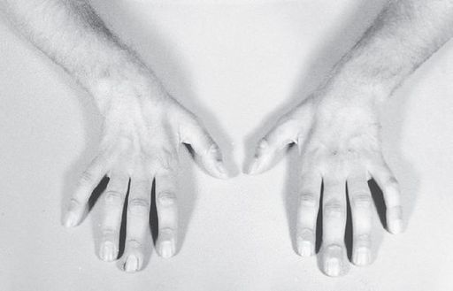
FIGURE 16.98
Wasting the small muscles of the hands. J Nicklin. Disorders of nerve II: polyneuropathies. In
Physical management in neurological rehabilitation
, 2nd edn. Fig 14.1. Mosby, Elsevier, 2004, with permission.
Causes
1.
Nerve lesions:
a.
median and ulnar nerve lesions
b.
brachial plexus lesions
c.
peripheral motor neuropathy (in the examination, don’t forget hereditary motor and sensory neuropathy).
2.
Anterior horn cell disease:
a.
motor neurone disease
b.
polio
c.
spinal muscular atrophies (e.g. Kugelberg-Welander disease).
3.
Myopathy:
a.
dystrophia myotonica – forearms more affected than the hands
b.
distal myopathy.
4.
Spinal cord lesions:
a.
syringomyelia
b.
cervical spondylosis with compression of C8 segment
c.
other (e.g. tumour).
5.
Trophic disorders:
a.
arthropathies (disuse)
b.
ischaemia, including vasculitis
c.
shoulder–hand syndrome.
HINT
When distinguishing an ulnar nerve lesion from a C8 root/lower trunk brachial plexus lesion, remember that the sensory loss of a C8 lesion extends proximal to the wrist and the thenar muscles are involved with a C8 root or lower trunk brachial plexus lesion. Distinguishing a C8 root from a lower trunk brachial plexus lesion is difficult clinically, but the presence of Horner’s syndrome or an axillary mass suggests that the brachial plexus is affected.
FEMORAL NERVE (L2, L3, L4) LESION
Clinical features
1.
Weakness of knee extension (quadriceps paralysis).
2.
Slight hip flexion weakness.
3.
Preserved adductor strength.
4.
Loss of knee jerk.
5.
Sensory loss involving the inner aspect of the thigh and leg.
SCIATIC NERVE (L4, L5, S1, S2) LESION
Clinical features
1.
Weakness of knee flexion (hamstrings involved).
2.
Loss of power of all muscles below the knee causing a foot drop, so the patient may be able to walk, but cannot stand on the toes or heels.
3.
Knee jerk intact.
4.
Loss of ankle jerk and plantar response.
5.
Sensory loss along the posterior thigh and total loss below the knee.
COMMON PERONEAL (LATERAL POPLITEAL) NERVE (L4, L5, S1) LESION
Clinical features
1.
Foot drop and loss of foot eversion only.
2.
Sensory loss (minimal) over the dorsum of the foot.
Note:
The reflexes are normal.
HINT
If there is a foot drop, test foot inversion and eversion.
Inversion
of the foot will be normal with a peroneal nerve compression but
absent
with a L5 radiculopathy. (Eversion is absent with both lesions.)
LATERAL CUTANEOUS NERVE OF THE THIGH LESION
Meralgia paraesthetica is caused by compression of this nerve, which may result in sensory loss and/or hyperaesthesia over the lateral aspect of the thigh, but no motor loss.
CAUSES OF FOOT DROP (
FIG 16.99
)
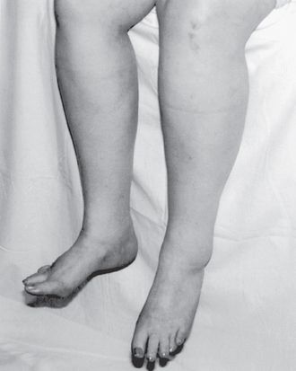
FIGURE 16.99
Foot drop in 45-year-old patient with amyotrophic lateral sclerosis. R B Daroff.
Bradley’s neurology in clinical practice
, 6th edn. Fig 74.5. Saunders, Elsevier, 2010, with permission.
1.
Common peroneal nerve palsy.
2.
Sciatic nerve palsy.
3.
Lumbosacral plexus lesion.
4.
L4, L5 root lesion.
5.
Peripheral motor neuropathy.
6.
Distal myopathy.
7.
Motor neurone disease.
8.
Precentral gyrus lesion.
HINT
Remember, if there is a foot drop, test the ankle jerk carefully. As a very rough rule of thumb, if it is absent, an S1 lesion should be suspected; if it is normal, a common peroneal palsy should be considered; if it is increased, an upper motor neurone lesion is likely.
Notes on spinal cord lesions
Assessment of the paraplegic patient
1.
Is there also a sensory level? Patterns of sensory loss depend on the level and type of lesion. Consider:
a.
cord compression, which causes a loss of all modalities bilaterally below the level involved (
note:
extrinsic compression may spare the perineum), and radicular pain and lower motor neurone weakness are present at the level of spinal compression
b.
transverse myelitis
c.
anterior spinal artery occlusion (posterior column function is spared)
d.
intrinsic cord lesion
e.
multiple sclerosis.
2.
Back examination: for example, deformity, tenderness or bruits may provide clues about the underlying disease process.
3.
Arm involvement? Consider:
a.
cervical spondylosis
b.
syringomyelia
c.
motor neurone disease
d.
multiple sclerosis.
4.
Cranial nerve lesions? Consider:
a.
motor neurone disease
b.
multiple sclerosis.
5.
Peripheral neuropathy? Consider:
a.
vitamin B
12
deficiency
b.
Friedreich’s ataxia
c.
carcinoma
d.
hereditary spastic paraplegia
e.
syphilis.
HINT
Intracranial lesions (e.g. parasagittal meningioma) cause paraplegia in extension only, whereas spinal cord lesions cause paraplegia in flexion or extension (i.e. flexor reflexes are released with spinal lesions).
Important motor and reflex changes of spinal cord and conus compression
Lower motor neurone signs occur at the level of the root lesion and upper motor neurone signs occur below the lesion.
UPPER CERVICAL
1.
Upper motor neurone signs in the upper and lower limbs.
2.
Paralysis of the diaphragm occurs with a lesion above C4.
C5
1.
Lower motor neurone weakness and wasting of the rhomboids, deltoids, biceps and brachioradialis.
2.
Upper motor neurone signs affect the rest of the upper and all the lower limbs.
3.
The biceps jerk is lost.
4.
The supinator jerk is ‘inverted’.
C8
1.
Lower motor neurone weakness and wasting of the intrinsic muscles of the hand.
2.
Upper motor neurone signs in the lower limbs.
Midthoracic
1.
Intercostal paralysis (cannot be detected clinically).
2.
Loss of upper abdominal reflexes at T7 and T8.
3.
Upper motor neurone signs in the lower limbs.
T10–T11
1.
Loss of the lower abdominal reflexes and upward displacement of the umbilicus on contraction (Beevor’s sign).
2.
Upper motor neurone signs in the lower limbs.
L1
1.
Cremasteric reflexes lost (normal abdominal reflexes).
2.
Upper motor neurone signs in the lower limbs.
L4
1.
Lower motor neurone weakness and wasting of the quadriceps.
2.
Knee jerk lost.
L5 and S1
1.
Lower motor neurone weakness of knee flexion and hip extension (S1) and abduction (L5), plus calf and foot muscles.
2.
Knee jerk present.
3.
No ankle jerk or plantar response.
4.
Anal reflex present.
S3–S4
1.
No anal reflex.
2.
Saddle sensory loss.
3.
Normal lower limbs.
HINT
Look for a urinary catheter if suspicious of a spinal cord lesion – in fact, always look for one.
Important syndromes
SUBACUTE COMBINED DEGENERATION OF THE CORD (VITAMIN B
12
DEFICIENCY)
Clinical features
1.
Symmetrical posterior column loss (vibration and position sense), causing an ataxic gait.
2.
Symmetrical upper motor neurone signs in the lower limbs with absent ankle reflexes; knee reflexes may be absent or, more often, exaggerated.
3.
Peripheral sensory neuropathy (less common and mild).
4.
Optic atrophy (occasionally).
5.
Dementia (occasionally).
The combination of upper motor neurone signs causing an extensor plantar response plus peripheral neuropathy causing loss of knee and ankle jerks is a distinctive pattern.
Causes of an extensor plantar response plus absent ankle jerk include: