i bc27f85be50b71b1 (116 page)
Read i bc27f85be50b71b1 Online
Authors: Unknown


VASCULAR SYSTEM AND HEMATOLOGY
379
• The presence, location, and severity of bone or joint pain using
an appropriate pain scale (See Appendix V I.)
• Joint range of motion and integrity, including the presence of
effusion or bony abnormality
• Presence, location, and intensity of parasthesia
• Blood pressure and heart rate for signs of hypovolemia (See Palpation in the Vascular Evaluation section for a description of viral sign changes with hypovolemia.)
Laboratory Studies
In addition to the history and physical examination, the clinical diagnosis of hematologic disorders is based primarily on laboratory studies.
Complete Blood Cell COLlllt
The standard complete blood cell (CBC) count consists of a red blood
cell (RBC) count, WBC count, WBC differential, hematocrit (Hct)
measurement, hemoglobin (Hgb) measurement, and platelet (Pit)
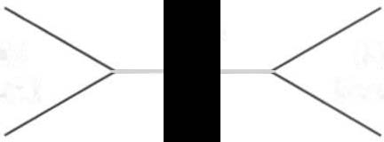
count. Table 6-7 summarizes the CBC. Figure 6-'1 illustrates a common method used by physicians to document portions of the CBC in daily progress notes. If a value is abnormal, it is usually circled within
this "sawhorse" figure.
Clinical Tip
• Hct is accurate in relation to fluid status; therefore, Hct
may be falsely high if the patient is dehydrated and falsely
low if the patient is fluid overloaded.
• Hct is approximately three times the Hgb value.
•
A low Hct may cause the patient to experience weakness, dyspnea, chills, or decreased activity tolerance, or it
may exacerbate angina.
• The term pancytopenia refers to a significant decrease
in RBCs, all types of WBCs, and Pits.
Erythrocyte Indices
RBC, Hct, and Hgb values are used to calculate three erythrocyte
indices: ( I ) mean corpuscular volume (MCV), (2) mean corpuscular
Hgb, and (3) mean corpuscular Hgb concentration. At mOst institu-

Table 6-7. Complete Blood Cell Count: Values and Interpretation-
w
00
o
Test
Description
Value
Indication/Inrerpreration
Red blood cell (RBC)
Number of RBCs per �I
Female: 3.8-5.1 million/�I To assess blood loss, anemia, polycythemia.
�
coum
of blood
Male: 4.3-5.7 million/�I
Elevated RBC count may increase risk of
�
venous stasis or thrombi formation.
i::
Increased: pol)'cythemia vera, dehydration,
J:
>
severe chronic obstructive pulmonary dis
�
ease, acute poisoning.
g
Decreased: anemia, leukemia, fluid overload,
'"
recent hemorrhage.
o
'"
White blood cell
Number of WBCs per �I
4.5-11.0xlOJ
To assess the presence of infection, inflamma
�
J:
(WBC) count
of blood
(4,500-11,000)
tion, allergens, bone marrow integrity.
-<
�
Monitors response to radiation or chemo-
�
therapy.
r
Increased: leukemia, infection. tissue necrosis.
i
Decreased: bone marrow suppression.
�
�
WBC differential
Proportion (%) of the
Neucrophils 54-62 %
To determine the presence of infectious
�
different types of
Lymphocytes 23-33%
states.
WBCs (out of 100
Detect and classify leukemia.
Monocytes 3-7%
cells)
Eosinophils 1-3%
Basophils 0-0.75%

Hematocrit (Het)
Percentage of RBCs in
Female: 35-40%
To assess blood loss and fluid balance.
whole blood
Male: 39-49%
Increased: Polycythemia, dehydration.
Decreased: anemia1 acute blood loss,
hemodilution.
Hemoglobin (Hgb)
Amount of hemoglobin
Female: 12-16 gllOO ml
To assess anemia, blood loss, bone marrow
in 100 ml of blood
Male: 13.5-17.5 gllOO 1111
suppression.
Increased: polycythemia, dehydration.
Decreased: anemia, recent hemorrhage, fluid
overload.
Platelets (Pit)
Number of platelets in
150-450 X 10'
To assess thrombocytopenia.
�I of blood
150,000-450,000 �I
Increased: polycythemia vera, splenectomy,
malignancy.
Decreased: anemia, hemolysis, DIC, ITP,
;;
viral infections, AlDS, splenomegaly, with
�
8
radiation or chemotherapy.
5:
"
·Lab values vary among laboratories. RBC, hemoglobin, and platelet values vary with age and gender.
�
AIDS = acquired immunodeficiency syndrome; DIC = disseminated intravascular coaguloparhy; ITP = idioparhic thrombocytopenic purpura.
Sources: Adapted from RJ Etin. Laboratory Reference Inrervals and Values. In L Goldman, JC Bennett (cds), Cecil Textbook of Medi
�
cine, Vol. 2 (21 Sf cd). Philadelphia: Saunders, 2000;2305; and E Marassarin-Jacobs. Assessment of Clienrs with Hemarologic Disorders.
�
In JM Black, E Matassarin-Jacobs, M,edical-Surgical Nursing Clinical Management for Continuity of Care (5rh ed). Philadelphia: Sauno ders, 1997;1465.
J:
� 5 C) -<
w
00
-


382 AClJTE CARE HANDBOOK FOR I)HVSICAL THERAPISTS
Hgb
WBC
Pit
Count
Count
Hct
Figure 6-1. Illustration of portions of the complete blood cell count in shorthand format. (Hcl ::: hematocrit; Hgb = hemoglobin; Pit = platelet; \lIBe =
white blood cell.)
tions, these indices are included in the CSc. Table 6-8 summari1.es
these indices.
Erythrocyte Sedimelltatio/1 Rate
The erythrocyte sedimentation rate, often referred to as the sed rate,
is a measurement of how fast RSCs fall in a sample of anticoagulated
blood. Normal values vary widely according to laboratory method.
The normal value is 1-20 mm per hour for men and 1-15 mm per
hour for women. The sedimentation rate is a nonspecific screening
tool used to determine the presence or stage of inflammation, or the
need for further medical testing, or it is used in correlation with the
clinical course of such diseases as rheumatoid arthritis or temporal
arteritis. II It may be elevated in systemic infection, collagen vascular
disease, and human immunodeficiency virus. Erythrocyte sedimentation rate may be decreased in the presence of sickle-cell disease, polycythemia, or liver disease or carcinoma.
Peripheral Blood Smear
A blood sample may be examined microscopically for alterations in
size and shape of the RSCs, WBCs, and Pits. RBCs are examined for