i bc27f85be50b71b1 (134 page)
Read i bc27f85be50b71b1 Online
Authors: Unknown
increasing or decreasing sweat production, superficial blood flow,
or both.
2.
Protection. The skin acts as a barrier to protect the body
from micro�organism invasion, ultraviolet (UV) radiation, abra�
sion, chemicals, and dehydration.
3.
Sensation. Multiple sensory cells within the skin detect
pain, temperature, and touch.
4.
Excretion. Heat, sweat, and water can be excreted from
the skin.
5.
Immunity. Normal periodic loss of epidermal cells removes
micro-organisms from the body surface, and immune cells in the
skin transport antigens from outside the body to the antibody cells
of the immune system.
6.
Blood reservoir. Large volumes of blood can be shunted
from rhe skin to central organs or muscles as needed.
7. Vitamin D synthesis. Modified cholesterol molecules ate
convened to vitamin 0 when exposed to UV radiation.


438 ACUTE CAR£ HANDBOOK FOR PHYSICAL TIII;RAJ'ISTS
Table 7-1. Normal Skin Layers: Structure and Function
Layer
Composition
Function
Epidermis
Strarum
Dead
Tough outer layer that protectS
corneum
keratinocytes
deeper layers of epidermis
Pigment layer
Melallocytes
Produces melanin to prevent
ultraviolet absorption
Stratum
Mature
Produces keratin to make the skin
granulosum
keratinocytes
waterproof
Langerhans' cells
Interacts with immune cells
Stratum
Keratinocytes
Undergoes mirosis to continue skin
spinosum
cell development but to a lesser
degree than basal
Stratum basale
ew keratinocytes
The origin of skin cells, which
undergoes mitosis, then moves
superficially
Merkel's cells
Detects touch
Dermis
Papillary layer
Areolar
Binds epidermis and dermis
connective
rogether
tissue
Meissner's
Detects light rouch
corpuscles
Blood and lymph
Provides circulation and drainage
vessels
Free nerve endings
Detects heat and pain
Reticular layer
Collagen, elastin,
Provides strength and resilience
and reticular
fibers
Hypodermis
Subcutaneous fat
Provides insulation and shock
absorption
Pacinian cells
Detects pressure
Free nerve endings
Detects cold
Source: Data from GA Thibodeau, KT Panon (cds), Structure and Function of the I�y
(11th ed). St Louis: Mosby, 2000.


BURNS AND WOUNDS 439
Pathophysiology of Burns
Skin and body tissue destrucrion occurs from rhe absorprion of heat
energy and results in tissue coagulation. This coagulation is depicted
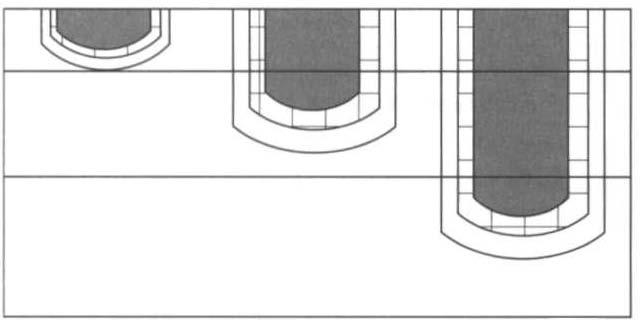
in zones (Figure 7-2). The ZOl1e of coagu/ali01l, locared in the cemer of
the burn, is the area of greatest damage and contains nonviable tissue
referred to as eschar. Alrhough eschar covers rhe surface and may
appear to take rhe place of skin, ir does nor have any of rhe characreristics or functions of normal skin. Instead, eschar is constrictive, attracts micro-organisms, and houses burn toxins that may circulate
throughout rhe body.' The Z01le of stasis, which surrounds rhe zone of
coagularion, comains marginally viable rissue. The Z01le of hyperemia, rhe ourermosr area, is the least damaged and heals rapidly.
Sequelae of Bum I1Ijury
A series of physiologic evems occurs afrer a burn (Figure 7-3). The
physical rherapisr musr appreciare rhe mulrisysrem effecrs of a
burn injury-namely, rhar rhe merabolic demands of the body
increase dramarically. Tissue damage or organ dysfuncrion can be
immediate or delayed, minor or severe, and local or systemic. A
A
B
c
Epidermis
Dermis
Subcutaneous
tissue
Figure 7-2. The ZOlles of coagulatioll. Superficial bum (AJ. Partial-thickness
bllm (8). Ful/-thickness bum (e). (Modified {rolll WG Wil/iallls, LG Phil/ips.
Pathophysiology of the Bum Wound. 111 DN Hemdo" {ed!. Total Burn Care.
London: Saunders, 1996;65,)


