i bc27f85be50b71b1 (64 page)
Read i bc27f85be50b71b1 Online
Authors: Unknown

l\.lUSCULOSKELETAL SYSTEM
205
Figure 3-18. Left total elbow arthroplasty shown ;n flexion.
lene articulation that connects with a locking pin or snap-fit deviceS!
and is indicated for use if there is injury to the musculature and ligamentous structures of the elbow. Implants are conStructed of materials similar to tho e of orher total joint arthroplasties, and fixation techniques vary with cemented, uncemented, and hybrid (a combination of cemented and uncemcnted) fixation designs. Patient selection, surgical
proficiency, and improved prosthetic design have improved outcomes
and reduced complications after TEA.
Complications after a TEA include component loosening (usually
the humeral component); joint instabiliry, including dislocation or
subluxation; ulnar nerve palsy; triceps weakness; delayed wound
healing; and infection.5o
Immediate rehabilitation of the patient with a TEA includes functional mobility training, ADL training, edema control, and ROM
exercises per surgeon protocol. If available, an elbow CPM may be
used. Passive elbow flexion and extension ROM may be allowed as
tolerated according to patient pain levels while strengthening exercises are avoided.


206
AClJrE CARE HANDBOOK FOR PHYSICAL THERAPISTS
Total Ankle Arthroplasty
Total ankle arthroplasty has had difficulty gaining populariry due
to i ncreased failure rare with early prosrheric designs. There has
been increased incidence of complications involving superficial and
deep wound infections, loosening of the prosthesi , and associared
subralar and midtarsal degenerative joint disease. Debilitating
arthritic pain is the primary indication for total ankle arthroplasty.
Currently, the total ankle arthroplasty i s optimal for an elderly
patient with low physical demands, normal vascular status, good
bone stock, and no immunosuppression.53 Progress has been made
with newer prosthetic designs and improved surgical techniques
involving limited periosteal stripping, meticulous arrention to positioning, and limited rigid internal fixation.
There are twO rypes of prostheric designs that have been developed to create a semiconstrained articulation. A two-component design with syndesmosis of the tibia and fibula consists of a tibial
articular surface that is wider than the talar component. Fibular
motion is eliminated, and a greater surface for fixation of the tibial
component is created.53 The second prosthetic design involves
three components: a flat metal tibial plate, a metal talar component, and a mobile polyethylene bearing H This system allows for multiplanar motion while maintaining congruency of the articulating surfaces. Both types of design consist of metal alloy and polyethylene components and rely on bony ingrowth for fixation.
Technical skill and experience in component alignment and
advanced instrumentation for component fixation arc important
aspects to be considered to improve postoperative outcomes. Current short-term results have shown reasonable increases in function, with little or no pain in appropriately selected patients.53
During the early postoperative period, care is taken to prevent
wound complications. A shorr leg splint is u sed until sutures are
removed, and the patient remains non weight bearing until bony
ingrowth is satisfactory (approximately 3 weeks w ith hydroxyapatite-coated implants).5J Early postoperative physical therapy treatment focuses on functional mobility and the patient's ability to maintain non weight bearing. The patient should be educated in
edema reduction and early non-weight-bearing exerci es, including
ankle ROM and hip and knee strengthening if prescribed by the
surgeon.

MUSCULOSKELETAL SYSTE.M
207
Total joint Arthroplasty lll{ection and Resecti01I
The percentage of rotal joint arthroplasties that become infected (septic)
is relatively small. However, a patient may present at any time after a
joint arthroplasty with fever, wound drainage, persistent pain, and
increased white blood cell count, erythrocyte sedimentation rate or Creactive protein.s, Infection is often diagnosed by aspirating the joint, culturing joint fluid specimens, and examining laboratory results from
the aspirate. Once the type of organism is identified, there are several different avenues to follow for treatment of the infection. Treatment choices include antibiotic treatment and suppression, debridement with prosthesis retention or removal, primary exchange arthroplasty, tvvo-stage reimplantation, or, in life-threatening instances, amputation.55
\'V'ithin the early postOperative period, an infection in soft tissue with
well-fixed implants can be treated with an irrigation and debridement
procedure, exchange of polyethylene components, and antibiotic treatment. With an infection penetrating the joint space to the cement-bone interface, it is necessary to perform a total joint arthroplasty resection,
consisting of the removal of prosthetic components, cement, and foreign material with debridement of surrounding tissue. A 6-week course of intravenous antibiotics should be followed before reimplantation of
joint components.'s The most successful results of deep peri prosthetic
infection have been obtained with the two-stage reirnplantation and
antibiotic treatment.45 With reimplantation of prosthetic components,
fixation depends on the quality of the bone, need for bone grafting, and
use of antibiotic-impregnated bone cemenr.43
Resection arthroplasty of the hip or knee is commonly seen in the
acute care orthopedic setting. A two-staged reimplantation is commonly performed. First, the hip or knee prosthetic is resected, and a cement spacer impregnated with antibiotics is placed in the joint to
maintain tissue length and allow for increased weight bearing. Then,
the prosthesis is re-implanted when the infection has c1eared.'s,ss If
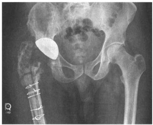
the eXTent of the infection does not allow for reimplantation of prosthetic components, a knee resection arthroplasty or hip Girdlestone (removal of the femoral head, as in Figure 3-19),55 may be rhe only
option fot the patient. Often, instability can be associated with these
surgeries, leading to decreased function, and the patient often requires
an assistive device for ambulation. Unlike a hip resection, a patient
with a knee resection may undergo a knee arthrodesis or fusion with
an intermedullaty nail to provide a stable and painless limb.

