Life on a Young Planet (21 page)
Read Life on a Young Planet Online
Authors: Andrew H. Knoll
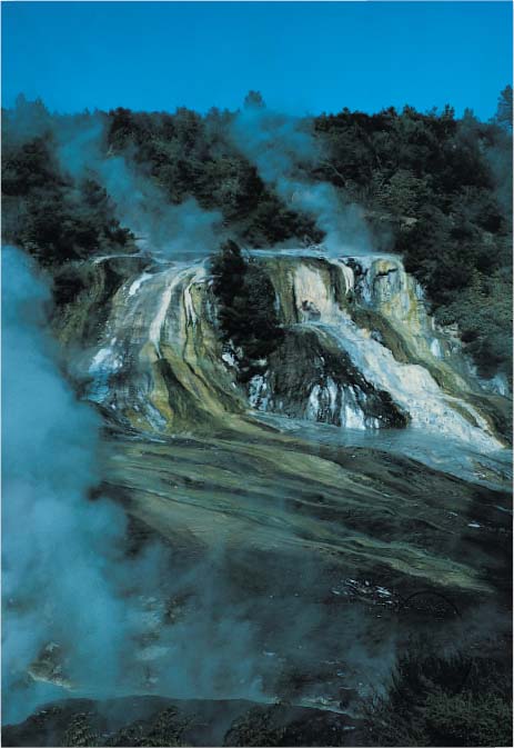
Plate 1.
Microbial ecosystem around a hot spring at Orakei Korako, New Zealand. The long blue-green streamers consist of cyanobacteria. Populated by metabolically diverse Bacteria and Archaea, modern hot springs suggest what life may have been like on the early Earth.
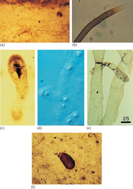
Plate 2.
Fossils in Akademikerbeen cherts and shales, and some living counterparts. (a) Filamentous fossils of mat-forming microorganisms in Spitsbergen chert; each tube is about 10 microns wide. (b) The cyanobacterial genus,
Lyngbya
, which provides a modern counterpart for the fossils in a. (The specimen is 15 microns wide.) Note the extracellular sheath that surrounds the ribbon of cells. Because it is not easily destroyed by bacteria, this sheath, rather than the cells it contains, is likely to enter the fossil record. (c)
Polybessurus bipartitus
, a distinctive stalk-forming microorganism in Spitsbergen cherts; specimen is about 35 microns wide. (d) A modern stalk-forming cyanobacterium that forms crusts on tidal flats of Andros Island, Bahamas; each specimen is 15 microns wide. This living counterpart to
Polybessurus
was discovered on the basis of environmental predictions made from the 600–800-million-year-old fossils. (e) Multicellular fossil from Spitsbergen shale comparable to the living green alga
Cladophora
(tubes 25 microns across). (f) Vase-shaped microfossils of tiny protozoans (fossil is 100 microns long; see
chapter 9
for further discussion). (Image (b) courtesy of John Bauld; (e) courtesy of Nicholas Butterfield)
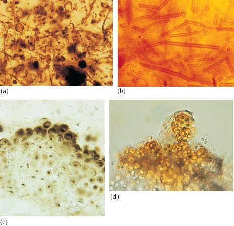
Plate 3.
Early Proterozoic microfossils and their modern counterparts. (a) A microscopic view of Gunflint chert, chockablock with tiny fossils. (b)
Leptothrix
, a modern iron-loving bacterium thought to be similar to the filaments in Gunflint fossil assemblages. In both figures, the filamentous organisms are 1–2 microns across. (c)
Eoentophysalis
cyanobacteria in early Proterozoic chert from the Belcher Islands, Canada. (d) A modern
Entophysalis
species for comparison (ellipsoidal envelopes around cells are 6–10 microns wide in both illustrations). (Photo (c) courtesy of Hans Hofmann; photo (d) courtesy of John Bauld)
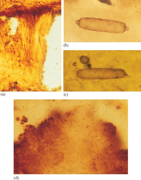
Plate 4.
Cyanobacterial microfossils in cherts of the 1.5-billion-year-old Bil’yakh Group. (a) A vertical tuft of tubular filaments, preserved in this orientation by very early formation of calcium carbonate cement (each filament is about 8 microns across). (b) A filamentous cyanobacterium, showing how cells were arranged along its length; the specimen is actually preserved as a lightly pigmented cast, originally made in rapidly cemented carbonate sediment (fossil is 85 microns long).(c)
Archaeoellipsoides
, the large (80 microns long, in this case) cigar-shaped fossil interpreted as the specialized reproductive cell of an
Anabaena
-like cyanobacterium. (d) 1.5-billion-year-old mat-building colony of
Eoentophysalis
; see plate 3d for its modern counterpart.
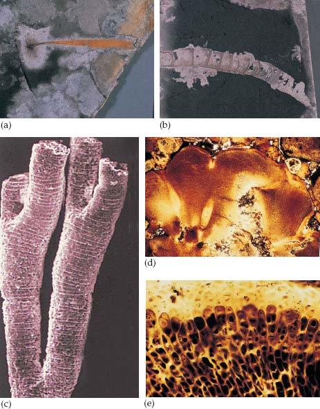
Plate 5.
Eukaryotic fossils in Doushantuo rocks. (a) Compression of a seaweed from shales at Miaohe; specimen is 2 inches long. (b) Tubular fossils of unknown but possibly animal origin, also from Miaohe; specimen is 3 inches long. (c) Small (150 microns across) branching tubes with distinctive cross-walls preserved in Doushantuo phosphates; possibly, these were made by early relatives of corals. (d) and (e), Multicellular red algae in Doushantuo phosphates; (d) illustrates a section through an alga, showing “cell fountains” and cylindrical recesses interpreted as reproductive structures; specimen is 1 millimeter across. Dark spots are individual cells; (e) shows well-preserved cells (6–10 microns in diameter) at higher magnification.
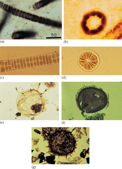
Plate 6.
Fossils of Proterozoic eukaryotes. (a) and (b) illustrate the fossil
Bangiomorpha
in ca. 1.2-billion-year-old cherts from arctic Canada. (c) and (d) show the living red alga
Bangia
. All specimens are about 60 microns in cross-sectional diameter. (e)
Tappania
, a 1.5-billion-year-old microfossil from northern Australia; fossil is 120 microns wide. (f) A lavishly ornamented microfossil (200 microns in diameter) interpreted as the reproductive spores of algae from ca. 1.3-billion-year-old rocks in China. (g) A large (more than 200 microns) spiny microfossil from ca. 570–590-million-year-old rocks in Australia. (Photos (a)–(d) courtesy of Nicholas Butterfield)
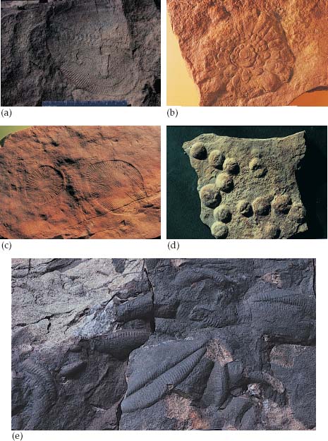
Plate 7.
Ediacaran fossils from Namibia and elsewhere. (a)
Swartpuntia
, a three-winged fossil found in the uppermost Proterozoic beds of the Nama Group; only two “wings” are evident in these fossils. (b)
Mawsonites
, a 4-inch disk from South Australia, interpreted an a sea anemone–like animal or the holdfast of a sea pen–like colony. (c)
Dickinsonia
, the most celebrated (and controversial) of vendobiont fossils. This specimen is from the Ediacara Hills of South Australia. (d)
Beltanelliformis
, a spherical green alga, here seen in latest Proterozoic sandstones from the Ukraine; specimens 1/2 to 3/4 inch across. (e)
Pteridinium
, another three-winged fossil found in sandstones of the Nama Group. (Photos (b) and (c) courtesy of Richard Jenkins)
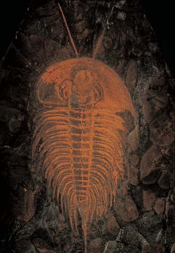
Plate 8.
The trilobite
Olenellus
, illustrating the tremendous complexity achieved by Early Cambrian animals. (Photo courtesy of Bruce Lieberman)
8 | The Origins of Eukaryotic Cells |