Examination Medicine: A Guide to Physician Training (89 page)
Read Examination Medicine: A Guide to Physician Training Online
Authors: Nicholas J. Talley,Simon O’connor
Tags: #Medical, #Internal Medicine, #Diagnosis
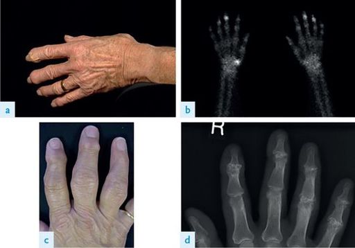
FIGURE 16.58
A 40-year-old patient with bilateral rheumatoid hand deformities. (a) Boutonnière deformities of left small, left ring, and right small fingers with simultaneous swan neck deformities of left long, right long, and right ring fingers. (b) Note inability to make a fist on the right hand (predominantly swan neck deformity) compared with the left hand (predominantly boutonnière deformity). (c) Radiograph. S J Sebastin, K C Chung. Reconstruction of digital deformities in rheumatoid arthritis.
Hand Clinics
, 2011. 27(1):87–104, Fig 1.
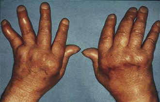
FIGURE 16.59
The hands of a patient with severe inflammatory arthritis, showing symmetrical deformity. J E Dacre, J G. Worrall. Rheumatology part 1 of 2: the rheumatological history.
Medicine
, 2010. 38(3):129–132, Fig 1.
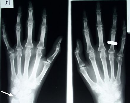
FIGURE 16.66
X-rays of the hands of a patient with early rheumatoid arthritis. Note the erosions of the metacarpal heads, reduced cartilage in the joint spaces and erosion of the ulnar styloid (arrow). Figure reproduced courtesy of The Canberra Hospital.
•
seronegative arthropathies – particularly psoriatic arthritis (see
Figs 16.60
,
16.61
,
16.68
and
16.69
)
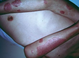
FIGURE 16.60
Psoriasis.
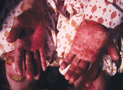
FIGURE 16.61
Pustular psoriasis.
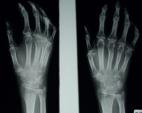
FIGURE 16.68
X-rays of the hands of a patient with polyarthritis secondary to connective tissue disease (CREST syndrome). There are destructive changes in all the joints (the DIP joints are not spared), and bony erosions are prominent. Figure reproduced courtesy of The Canberra Hospital.
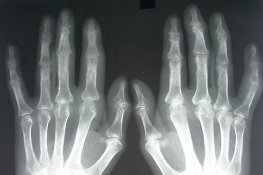
FIGURE 16.69
X-ray of the hands of a patient with psoriatic arthritis. Note bone erosion, loss of joint space and ‘pencil in cup deformity’ of the PIP joints. Figure reproduced courtesy of The Canberra Hospital.
•
polyarticular gout (look for tophi) (
Figs 16.62a, b
and
16.70
) or pseudogout (see
Fig 16.63
)
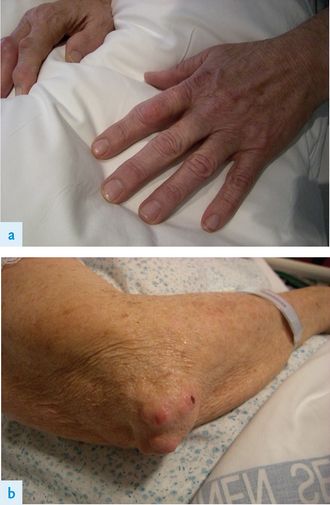
FIGURE 16.62
(a) and (b) Tophaceous gout.
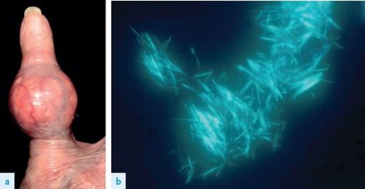
FIGURE 16.63
(a) Pseudogout. The swollen interphalangeal joint. (b) Calcium pyrophosphate crystals. A Alexandroff, N Kirkham, N Nayak.
The Lancet
. Fig 1a. 371(9618):1114. Elsevier, 2008, with permission.
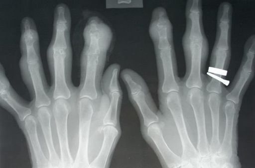
FIGURE 16.70
X-ray of the hands of a patient with severe gouty arthritis. Note the large soft-tissue masses and severe joint destruction. Figure reproduced courtesy of The Canberra Hospital.
•
primary generalised osteoarthritis (where DIP and PIP joint involvement is common) (see
Figs 16.64
,
16.65
and
16.71
).
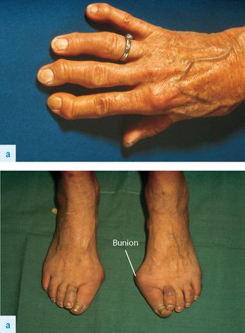
FIGURE 16.64
(a) and (b) Primary generalised osteoarthritis. N Talley, S O’Connor,
Clinical examination
, 7th edn. Fig 24.5a and b. Elsevier Australia, 2013, with permission.
Other books
A Lady's Guide to Improper Behavior by Suzanne Enoch
Other People's Heroes (The Heroes of Siegel City) by Petit, Blake M.
Casi un objeto by José Saramago
Flora's Defiance by Lynne Graham
Murder.com by Christopher Berry-Dee, Steven Morris
Claire De Lune by Christine Johnson
Surfacing (Spark Saga) by Melissa Dereberry
Athena by John Banville
Dream Horse by Bonnie Bryant
The Sword of Shannara Trilogy by Terry Brooks